

VARICOCELE
Varicocele, also known as varicose veins of the testicles, refers to the enlargement of veins located in the scrotum, which is the protective skin sac surrounding the testicles. It occurs in approximately 15% of the male population, and about 40% of infertile men (those experiencing male infertility) are found to have varicoceles. Varicoceles are rare in children under the age of 10.

Symptoms of Varicocele
The clinical presentation of varicoceles can vary. Some individuals with varicoceles may not experience any symptoms, while others may exhibit the following:
- Scrotal mass or swelling
- Scrotal pain
- Testicular atrophy (shrinkage)
- Infertility
- Subfertile conditions, which refer to situations where pregnancy is still possible.
Normally, the testicular veins (pampiniform veins) play a crucial role in sperm production and help regulate the testicular temperature, which should ideally be maintained at 35 degrees Celsius. Varicoceles disrupt this balance, causing the temperature to rise to 37 degrees Celsius. Varicoceles can be classified as primary or secondary.
Many cases of varicoceles are primary and result from congenital absence or insufficiency of valves in the testicular veins (internal spermatic veins). The left testicle is more commonly affected than the right, with an 85% occurrence on the left side. Bilateral varicoceles are not uncommon, accounting for 5% of cases. However, isolated right-sided varicoceles are extremely rare and warrant careful investigation into secondary causes.
Secondary varicoceles occur less frequently. They can develop due to external compression (e.g., lymph node), obstruction (e.g., renal vein obstruction), or the presence of a shunt between the kidney and spleen, leading to increased pressure in the testicular veins.

Diagnosis of Varicocele
Varicocele, also known as the dilation of the testicular veins, is typically more common on the left side of the testicles. Ultrasonography is a commonly used imaging method for diagnosing varicoceles. Here are the details based on ultrasound findings:
- Dilation of Testicular Veins (Pampiniform Plexus):
- There is dilation observed in the testicular veins (pampiniform plexus).
- These veins typically have a serpentine appearance.
- Their diameter is wider than 2-3 mm.
- Doppler Ultrasonography:
- Doppler ultrasonography is used to determine the degree of reflux (leakage).
- When breath is held and strain is applied, the flow is seen to reverse.
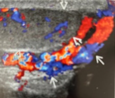
Doppler ultrasonography show dilateted testicular veins around testis.
- Venography:
- Venography is considered the gold standard for diagnosing varicoceles.
- This procedure is usually performed during varicocele embolization.
- A catheter is inserted into the testicular vein through the groin, and contrast material is injected.
- Findings:- Enlarged testicular veins are visualized.
- The retrograde flow of the contrast agent toward the scrotum (testicular sac) is demonstrated during venography.
- Magnetic Resonance Imaging (MRI):
- MRI is also used for diagnosis.
- In MR imaging, the dilated veins are depicted.
These details cover the fundamental findings used in the diagnosis of varicoceles. If you suspect a varicocele, it’s important to consult a healthcare professional for further evaluation.
Embolization in Varicocele Treatment
In varicocele treatment, there are primarily two methods: surgical intervention or embolization performed by interventional radiology. Varicocele is one of the surgically correctable causes of male infertility. Surgical ligation of the testicular veins is a treatment method.
In cases where varicocele recurs after surgery or when surgery is not desired, embolization of the testicular veins is applied. Varicocele embolization is performed in interventional radiology departments within angiography units. It is a same-day procedure that does not require general anesthesia.
Thin catheters called “kateters” are selectively inserted into the testicular veins through the groin or neck veins. Initially, contrast material is injected through these catheters to perform venography. During venography, the retrograde flow toward the testicular sac is demonstrated in the testicular veins. After this stage, embolization is carried out using coils, sclerosing agents, or adhesives known as “glue.”
The risks associated with varicocele embolization are rare. During embolization, there may be perforation and subsequent bleeding in the veins, as well as the possibility of small metallic coils (referred to as “koil”) migrating to the lungs.